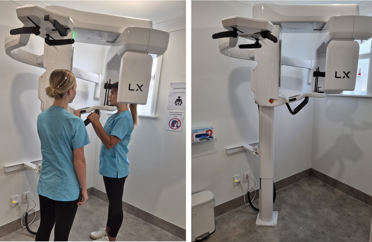
Cone Beam Computed Tomography (CBCT)
What is CBCT?
CBCT scans are a variation of traditional computed tomography (CT) scans.
The CBCT systems used by dental professionals rotate around the patient, capturing data using a cone-shaped X-ray beam.
Depending on the size and type of scan these data are used to reconstruct a three-dimensional (3D) image of the teeth, mouth, jaws and base of the sinuses.
What CBCT machine do you use?
The Dexis OP3D LX, with a resolution of 80 microns for the size of the voxel (the 3D equivalent of a pixel). This is one of the smallest resolutions available and ideal for endodontic diagnosis and treatment planning.
For more information see:
(https://dexis.com/en-us/op-3d-lx#features)
What are the advantages of CBCT scans over traditional radiography?
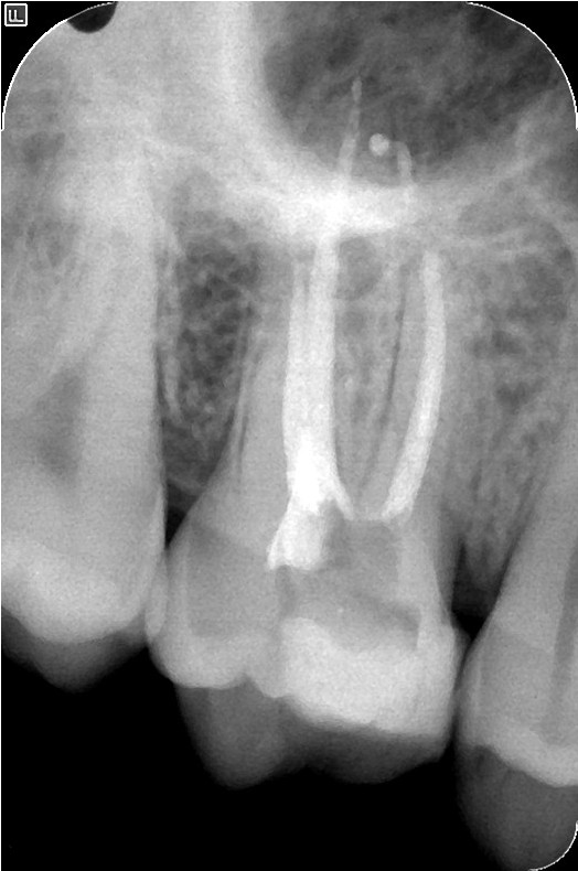
Traditional radiographs involve superimposition of a number of anatomical structures (which of course are 3-D) onto a 2-D image/film.
While at times this is sufficient in order to make a diagnosis, other times this is simply not clear enough.
CBCT scans are 3-D and so far more accurate in showing potential pathology associated with teeth and jaws.
Below we see two images: the first is of a traditional radiograph with some pathology visible around the roots of an upper molar, and the second is an image showing 2D slices of pathology visible around the entire tooth and sinus.
The actual 3-D image can be manipulated to show any combination of slices so one can "move through the tooth" to check for potential issues.
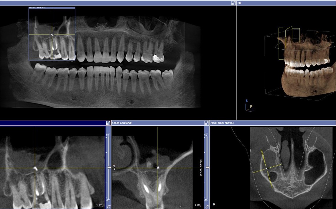
Studies have shown that a 3-D scan can change a diagnosis and/or treatment plan in around 50% of cases.
(https://pubmed.ncbi.nlm.nih.gov/25070420/ ; https://pubmed.ncbi.nlm.nih.gov/32628966/)
How does CBCT apply in Endodontics?
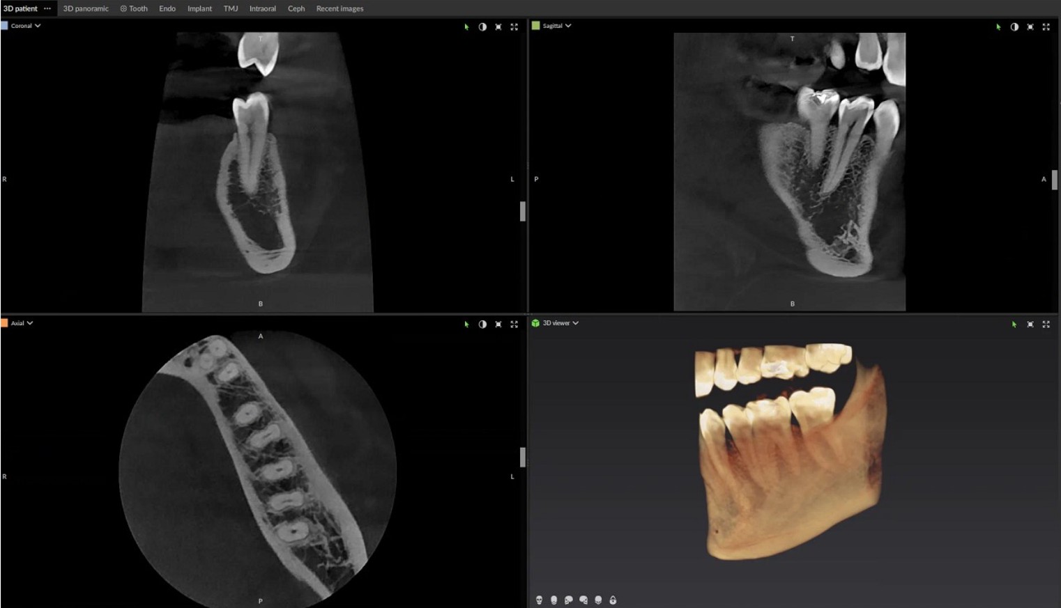
Helping to formulate a diagnosis of pulpal & periapical status, checking for extra/missed canals & roots, size & extent of resorptive lesions, surgery planning, trauma & potential fractures (depending on the type of fracture), degree of intrusion/extrusion, root development, and differential diagnosis of pain (tooth vs other sources) are some of the possible applications of CBCT in endodontics.
Below are some 2D slices of a CBCT scan.
What about the radiation level?
The radiation doses from dental CBCT exams are generally lower than other CT exams, but dental CBCT exams typically deliver more radiation than conventional dental X-ray exams. Generally a scan will be a small field of view (FOV) scan to focus on the area of interest at the highest possible resolution. This delivers about the same radiation as the increase in cosmic radiation received when flying from Auckland to Singapore return. Where a full mouth scan is needed (such as to try and reach a differential diagnosis for pain, or if a full mouth rehabilitation is involved) the radiation level is about the same as flying from Auckland to Europe return. This is less than a traditional chest X-ray using conventional film.
Generally we try and avoid CBCT scans during the first trimester of pregnancy. Please advise us if you are pregnant.
